
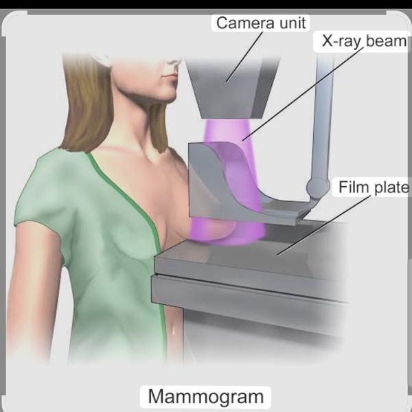
# Mammography in Rohini
2026-06-07T09:45:53  read more
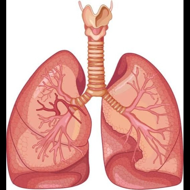

Chest ultrasound in rohini delhi Dr. Mittal Diagnostic Centre C-8/193 sector 8 Rohini Delhi 9811552279 7827070020 A chest ultrasound, or transthoracic ultrasonography, is a non-invasive imaging test that uses sound waves to create real-time images of the lungs, heart, pleural space, and other structures within the chest. A transducer is placed on the skin to send sound waves that bounce off internal structures and return as echoes, which are then translated into images. It's a useful tool for detecting pneumothorax, pleural effusion (fluid around the lungs), pulmonary consolidation (like pneumonia), and for guiding procedures such as thoracentesis (fluid removal). How it Works 1. Sound Waves: A handheld device called a transducer sends high-frequency sound waves into the body. 2. Echoes: These sound waves reflect off different tissues and structures within the chest, creating echoes. 3. Image Formation: The transducer captures these returning echoes, which are then processed by a computer to generate a live, moving black-and-white image on a monitor. What It Detects Chest ultrasound can identify various abnormalities, including: Pneumothorax: Air in the space between the lung and chest wall. Pleural Effusion: An abnormal buildup of fluid in the pleural space. Pulmonary Consolidation: Dense areas in the lung, often indicative of pneumonia. Interstitial Syndrome: Conditions like pulmonary edema (fluid in the lung tissue). Diaphragm Movement: How well the diaphragm is functioning. Uses Diagnosis: Rapid diagnosis of conditions like pneumonia, air in the chest, or fluid buildup. Guidance for Procedures: Guiding needles for biopsies or to remove fluid, making these procedures safer and more effective. Monitoring: Tracking changes in lung conditions over time. Point-of-Care Tool: Used directly at the patient's bedside in intensive care units and emergency departments, avoiding the need to move patients or wait for other tests. Advantages No Ionizing Radiation: Unlike X-rays or CT scans, it does not use harmful radiation. Real-time Imaging: Provides immediate, live views of chest structures. Non-Invasive: It's a simple, safe procedure performed on the skin's surface. Portable: Can be performed at the patient's bedside.
We hate spam too.
