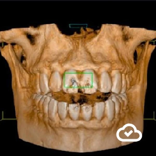
Best Dental X-rays and CBCT in Rohini
2026-05-14T17:52:57  read more
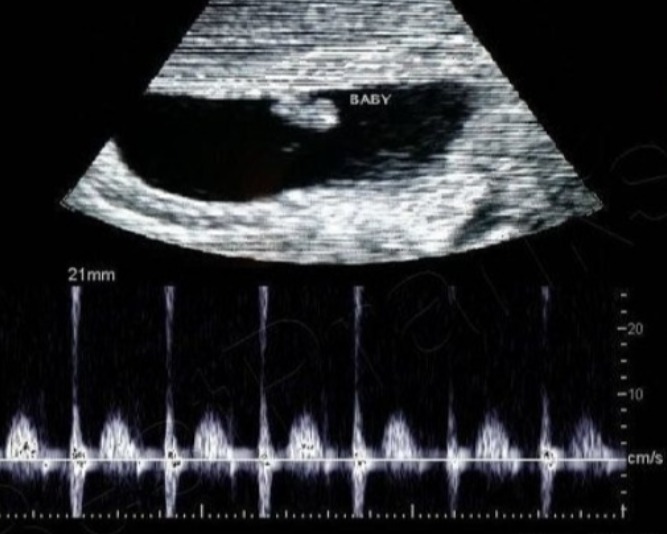

Early pregnancy ultrasound in rohini C-8/193 sec 8 rohini delhi 9811552279 7827070020 An early pregnancy ultrasound is a diagnostic tool used to confirm pregnancy, check for viability, and estimate the gestational age. Typically performed between 4 and 6 weeks, a transvaginal ultrasound is often clearer than an abdominal one at this stage and can help rule out ectopic pregnancies and other complications. A transvaginal ultrasound can visualize the gestational sac, yolk sac, and fetal pole, and later, the heartbeat and crown-rump length (CRL), which is the most accurate way to date the pregnancy. What an early pregnancy ultrasound checks for Confirmation of pregnancy: Confirms that the pregnancy is located in the uterus and not an ectopic pregnancy. Viability: Detects a fetal heartbeat to confirm the pregnancy is progressing normally. Gestational age: Provides an estimated number of weeks by measuring the size of the gestational sac, yolk sac, or embryo (CRL). Number of fetuses: Identifies if it is a single or multiple pregnancy. Other issues: Can detect internal bleeding or other complications. What to expect Transvaginal vs. abdominal: A transvaginal ultrasound is often preferred in early pregnancy because the transducer is placed in the vagina, providing clearer and more detailed images closer to the uterus and ovaries. An abdominal ultrasound may be used from around 7 weeks onwards. What is visible: As early as 4.5 weeks, the gestational sac can be seen. The yolk sac appears around 5 weeks, and the embryo and its heartbeat can often be detected by 6–8 weeks. Accuracy: Measuring the crown-rump length (CRL) is the most accurate method for dating the pregnancy, with accuracy up to plus or minus 3 to 5 days. Duration: A scan typically takes 20 to 30 minutes, but can take longer if a clear view is difficult to obtain.
We hate spam too.