

+918048036246

Currently it only shows your basic business info. Start adding relevant business details such as description, images and products or services to gain your customers attention by using Boost 360 android app / iOS App / web portal.
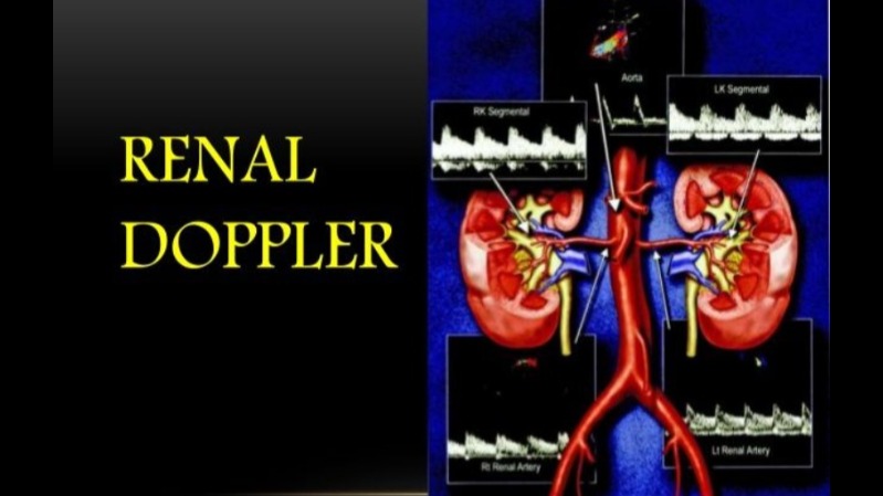
Renal Doppler ultrasound in rohini delhi c-8/193 sec8 rohini delhi 9811552279 7827070020 A Renal Doppler test is a non-invasive ultrasound used to assess blood flow within the renal arteries and kidneys, helping diagnose conditions like renal artery stenosis (narrowing of the artery), chronic kidney disease, and transplant rejection. The procedure involves applying gel to the skin and using a transducer to send sound waves into the body, creating real-time images of blood flow and providing information about its speed and direction. The test is generally quick, takes about 30 minutes, and requires minimal preparation, although some specialists may recommend an empty stomach or fasting. Purpose The primary goal of a Renal Doppler is to: Detect renal artery stenosis (RAS): Identify narrowing in the arteries supplying blood to the kidneys, often a cause of uncontrolled high blood pressure. Evaluate Chronic Kidney Disease (CKD): Assess vascular changes in the kidneys to predict disease progression and help manage therapy. Monitor Transplanted Kidneys: Detect signs of rejection or other vascular issues in transplanted organs. Diagnose other renal conditions: Aid in identifying blockages, vascular changes, and other anomalies affecting the kidneys. Procedure Preparation: Typically, you will be asked to wear comfortable clothing. Some sources suggest fasting for a few hours before the test, while others advise against it, so it's best to follow specific instructions from your doctor or the clinic. Gel application: A small amount of gel is applied to the skin over the kidney area to ensure good contact between the transducer and the skin. Transducer placement: The technologist places the transducer against the skin. Image creation: The transducer emits high-frequency sound waves that travel through the body and bounce back as echoes. A computer uses these echoes to create real-time images of the blood flow in the renal arteries on a monitor. What to expect Non-invasive: The test is painless and does not involve any radiation. Real-time results: Images and blood flow patterns are viewed immediately on a video monitor. Speed: The scan itself usually takes less than 30 minutes to complete. Operator-dependent: The quality of the results is highly dependent on the skill of the sonographer. Key Parameters Measured Peak Systolic Velocity (PSV): The highest speed of blood flow during the heart's contraction. Resistive Index (RI): A measure of the resistance to blood flow within the kidney's smaller arteries. Renal Aortic Ratio (RAR): The ratio of the PSV in the renal artery to the PSV in the aorta. Flow patterns: Waveform analysis can identify normal, accelerated, or abnormal blood flow.
