

+918048036246

Currently it only shows your basic business info. Start adding relevant business details such as description, images and products or services to gain your customers attention by using Boost 360 android app / iOS App / web portal.
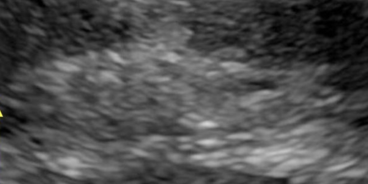
Soft tissue ultrasound near me C-8/193 sector 8 rohini delhi 9811552279, 7827070020 A soft tissue ultrasound uses high-frequency sound waves to create real-time images of superficial body structures, helping to diagnose conditions like skin and soft tissue infections, abscesses, foreign bodies, and soft tissue masses or lumps. This non-invasive procedure provides detailed views of the skin, fat, muscle, tendons, ligaments, nerves, and fascia, allowing clinicians to assess abnormalities, guide procedures like biopsies, and monitor conditions that may not be apparent through a physical examination alone. What it is and how it works High-frequency sound waves: An ultrasound probe (transducer) is moved over the skin, emitting high-frequency sound waves that bounce off tissues. Image creation: These echoes are captured by the probe and processed by a computer to generate real-time images displayed on a screen. Dynamic assessment: The ability to visualize tissues in motion allows for a dynamic assessment of conditions that may not be obvious with other imaging methods. What can be visualized Superficial structures: The epidermis, dermis, and subcutaneous fat are clearly visible, with different layers having distinct appearances. Underlying structures: The scan can also image fascia, muscle fascicles, tendons, ligaments, and nerves. Hard vs. soft tissues: Bone appears as a dense, bright white structure with dark shadowing behind it, while softer tissues have different densities and textures. Why it's used Infections: Helps to differentiate between cellulitis and an abscess, determine the size and depth of an infection, and guide drainage procedures. Lumps and masses: Used to evaluate palpable soft tissue masses, lumps, or bumps, and to distinguish between different types of lesions like cysts. Foreign bodies: Can help to locate and identify foreign bodies within the soft tissues. Injury assessment: Used to assess injuries to muscles, tendons, and ligaments. Point-of-care ultrasound (POCUS): A valuable tool for clinicians to use at the patient's bedside when a physical exam is inconclusive. Guided procedures: Provides guidance for procedures such as biopsies, helping to ensure accuracy and identify high-yield areas for tissue sampling.
