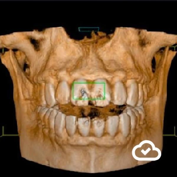
Best Dental X-rays and CBCT in Rohini
2026-05-14T17:52:57  read more
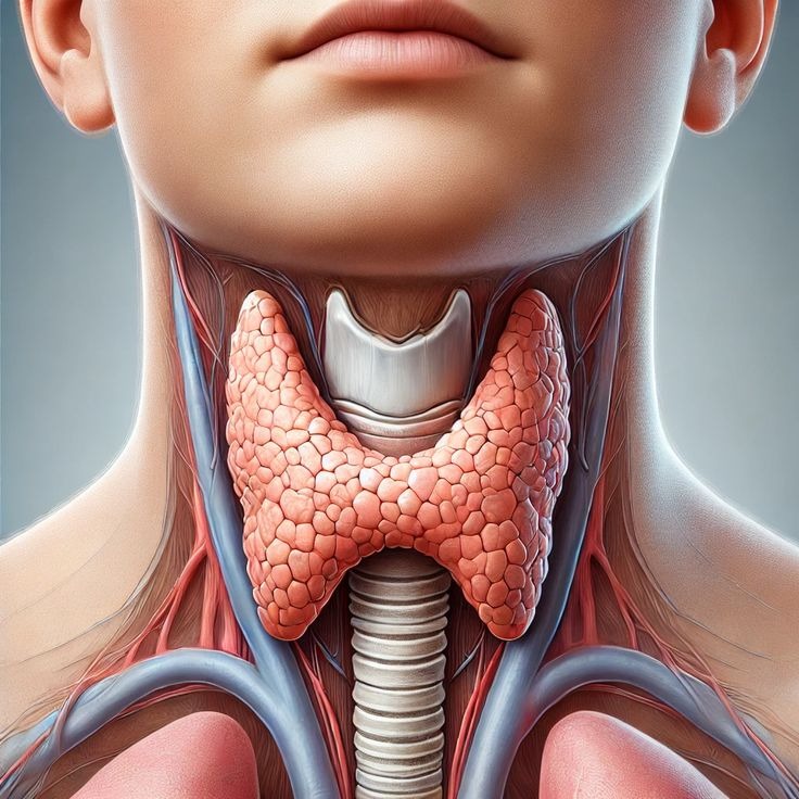

Thyroid ultrasound in rohini delhi thyroid ultrasound in delhi Dr. Mittal's Diagnostic Centre C-8/193 sector 8 rohini delhi 9811552279 7827070020 A thyroid ultrasound uses sound waves to create images of the thyroid gland in the neck, allowing doctors to evaluate nodules, cysts, goiters, and other abnormalities. The painless, non-invasive procedure involves applying a water-based gel and a handheld transducer to the neck to capture images on a monitor. It's a crucial tool for detecting abnormalities, characterizing suspicious nodules, and guiding procedures like fine-needle aspiration (FNA), but involves no radiation or side effects. What is a thyroid ultrasound? It's an imaging technique that uses high-frequency sound waves to produce detailed images of the thyroid gland, a gland that regulates metabolism. The images show the size, location, and characteristics of any nodules, cysts, or other structures within the gland. Why is it performed? Detection of Nodules: It's the most sensitive method for detecting thyroid nodules, which can be felt during a physical exam or found on other imaging tests. Evaluation of Abnormalities: It helps diagnose tumors, cysts, and goiters (enlarged thyroid). Cancer Detection: It is used in the detection and assessment of thyroid cancer and can help identify suspicious features associated with malignancy. Biopsy Guidance: The ultrasound can guide a fine-needle aspiration (FNA) biopsy, where a thin needle is used to take a sample of cells for examination. Monitoring: It helps monitor patients after treatment for early detection of tumor recurrence. What to expect during the procedure: 1. Preparation: Little to no special preparation is needed; you can eat and drink as usual. Remove jewelry from the neck area. 2. Positioning: You will lie face-up on an exam table, sometimes with a pillow under your neck to tilt your head back. 3. Gel Application: A small amount of water-based gel is applied to your neck to help the transducer make good contact. 4. Transducer Use: The technician will press a handheld wand (transducer) over the gel, moving it around the thyroid area. 5. Imaging: The transducer sends sound waves into your neck, and the returning echoes are translated into a 2D image on a monitor. 6. Completion: After the images are captured, the gel is wiped off your neck. The entire process typically takes about 30 minutes.
We hate spam too.